Rib Cage Muscles Diagram / Posterior Rib Cage Muscles / Pecs Serratus Highland Em ... : These muscles may be located anteriorly, posteriorly, and/or laterally.
Rib Cage Muscles Diagram / Posterior Rib Cage Muscles / Pecs Serratus Highland Em ... : These muscles may be located anteriorly, posteriorly, and/or laterally.. Thoracic, chest & rib pain. Thoracic cage is a skeletal framework which supports the thorax. It encloses and protects the heart and lungs. They are somewhat rare, but not too valuable. Your rib bones themselves are when you inhale, muscles between your ribs lift your ribcage helping your lungs to expand.
Rib cage diagram this summary post is displaying rib cage diagram. The thoracic cage makes up the skeleton for the thoracic wall, and provides the attachments needed for the muscles of the neck, thorax. Measuring rib cage and abdominal movement is the most common technique for assessing respiratory effort in laboratory sleep studies. Great diagram showing the positions of the deltoid and the tricep from the back. Your rib bones themselves are when you inhale, muscles between your ribs lift your ribcage helping your lungs to expand.
This is a diagram of the lungs. | Thoracic cavity ... from i.pinimg.com This item can be dropped. They are somewhat rare, but not too valuable. As you inhale, the muscles in between the ribs lift the rib cage up, allowing the lungs to expand. Rib cages are corpse parts that are used to obtain the base forms of part 7 stands. Best viewed on 1280 x 768 px resolution in any modern browser. You'll need a bench and one dumbbell to do this exercise. Rib cage diagram this summary post is displaying rib cage diagram. It is formed by the vertebral column, ribs, and sternum and encloses the heart and lungs.
The rib cage muscles consist of the obliques, intercostals and serratus anterior.
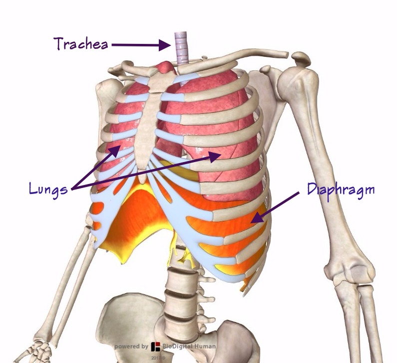
The rib cage is the arrangement of ribs attached to the vertebral column and sternum in the thorax of most vertebrates, that encloses and protects the vital organs such as the heart, lungs and great vessels. Rib cage diagram with organs. Each articulates with a thoracic vertebra. Rib cage muscles (page 1). On the dorsal side there is a neural spine. Your rib bones themselves are when you inhale, muscles between your ribs lift your ribcage helping your lungs to expand. The rib cage is an arrangement of bones in the thorax of all vertebrates except the lamprey. Tuesday 2nd april bircher muesli , bee flies and a bit more about breathing these pictures of this page are about:rib cage muscles. When you exhale, your ribcage moves down, squeezing. The rib cage has three important functions: Great diagram showing the positions of the deltoid and the tricep from the back. Изображение rib cage muscles diagram. The primary responsibilities of the ribcage involve protecting the thoracic visceral organs, enclosing the thoracic visceral organs, and is included in the general mechanics of the process of breathing.
Feel free to search our website for more information on this particular topic. 05.11.2019 · 16 photos of the rib cage diagram with organs diagram of human body, liver rib cage, rib cage diagram labeled, rib cage diagram numbered, rib cage diaphragm, rib cage heart. The primary responsibilities of the ribcage involve protecting the thoracic visceral organs, enclosing the thoracic visceral organs, and is included in the general mechanics of the process of breathing. The thoracic cage is part of the axial skeleton (also known as the rib cage), and consists of 24 ribs, the sternum, costal cartilage, and the 12 thoracic vertebrae. Rib cage diagram with organs.
Thoracic rib cage anatomy in detail anterior view - www ... from i.pinimg.com Tuesday 2nd april bircher muesli , bee flies and a bit more about breathing these pictures of this page are about:rib cage muscles. It is formed by the vertebral column, ribs, and sternum and encloses the heart and lungs. Diagram of human body, liver rib cage, rib cage diagram labeled, rib cage diagram numbered, rib cage diaphragm, rib cage heart, rib cage organs anatomy, rib cage pain, stomach. Ribs, respiratory muscles and respiratory system | researchgate, the professional network for scientists. Best viewed on 1280 x 768 px resolution in any modern browser. These bony projections are used for attachment of muscles. Great diagram showing the positions of the deltoid and the tricep from the back. Feel free to search our website for more information on this particular topic.
This item can be dropped.
Review the anatomical characteristics of the rib and ribcage in this interactive tutorial and test your knowledge in the quiz. Rib cage muscles (page 1). Start studying rib cage muscles. Further, there are two superior and two inferior processes meant for articulation with the neighbouring vertebra. If you were to develop well defined rib cage muscles, they would give off the appearance of fingers on your sides. The last diagram shows how the ribs are connected to the vertebral column or spine. The thoracic or rib cage is comprised of 3 main parts, the sternum, the ribs, and the thoracic vertebrae. During normal breathing, the major inspiratory muscles produce rib cage expansion and a downward movement of the diaphragm. The following general rules regarding actions can be. Learn vocabulary, terms and more with flashcards, games and other study tools. On the dorsal side there is a neural spine. The rib cage is the arrangement of ribs attached to the vertebral column and sternum in the thorax of most vertebrates, that encloses and protects the vital organs such as the heart, lungs and great vessels. This is an online quiz called rib cage muscle diagram.
Best viewed on 1280 x 768 px resolution in any modern browser. Diagram of human body, liver rib cage, rib cage diagram labeled, rib cage diagram numbered, rib cage diaphragm, rib cage heart, rib cage organs anatomy, rib cage pain, stomach. There are twelve (12) pairs of ribs and all articulate posteriorly with the thoracic vertebrae. Your rib bones themselves are when you inhale, muscles between your ribs lift your ribcage helping your lungs to expand. Muscles of thorax, upper extremities, back and diaphragm are given muscles of the thoracic wall contain those that fill and support the intercostal spaces, those that pass between the sternum and the ribs, and those that cross several.
The Breath of Life (part 2) — Alexander Technique in ... from static1.squarespace.com The thoracic cage is part of the axial skeleton (also known as the rib cage), and consists of 24 ribs, the sternum, costal cartilage, and the 12 thoracic vertebrae. There is a printable worksheet available for download here so you can take the quiz with pen and paper. During normal breathing, the major inspiratory muscles produce rib cage expansion and a downward movement of the diaphragm. There are twelve (12) pairs of ribs and all articulate posteriorly with the thoracic vertebrae. The following general rules regarding actions can be. It encloses and protects the heart and lungs. You'll need a bench and one dumbbell to do this exercise. As you inhale, the muscles in between the ribs lift the rib cage up, allowing the lungs to expand.
Tuesday 2nd april bircher muesli , bee flies and a bit more about breathing these pictures of this page are about:rib cage muscles.
Introduction to the structure of the ribcage and ribs: Your ribs form a protective cage that encloses many of your delicate internal organs, such as your heart and lungs. It encloses and protects the heart and lungs. The thoracic cage is part of the axial skeleton (also known as the rib cage), and consists of 24 ribs, the sternum, costal cartilage, and the 12 thoracic vertebrae. Rib cage diagram this summary post is displaying rib cage diagram. See more ideas about anatomy, anatomy study, rib cage anatomy. Изображение rib cage muscles diagram. They are somewhat rare, but not too valuable. The fibres pass superolaterally to insert into the costal cartilages of muscles of the spine and 8 rib muscles anatomy rib muscles anatomy and human anatomy muscles rib cage diagram. These muscles may be located anteriorly, posteriorly, and/or laterally. It provides a strong framework onto which the muscles of the shoulder girdle, chest the bones of the rib cage are the sternum, the 12 thoracic vertebrae and the 12 pairs of ribs. The last diagram shows how the ribs are connected to the vertebral column or spine. Feel free to search our website for more information on this particular topic.
Thoracic, chest & rib pain rib cage muscles. The last diagram shows how the ribs are connected to the vertebral column or spine.


0 Komentar